
Precision Imaging Starts with Distortion Control
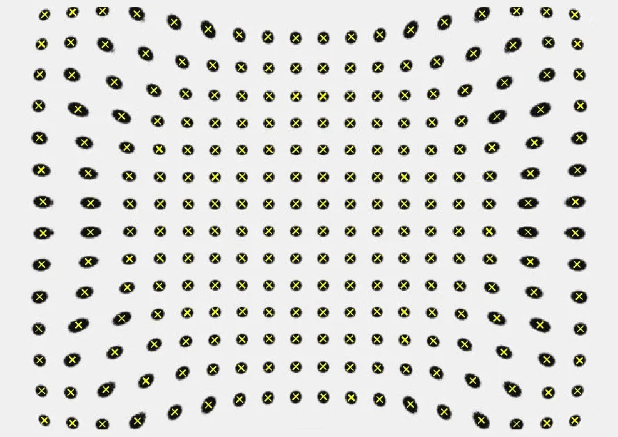
In medical and endoscopy imaging, every pixel matters. Image distortion, where straight lines appear curved or deformed, can directly impact diagnostic accuracy and procedural confidence. Ensuring geometrically precise images is therefore critical for modern medical camera systems.
Distortion in Medical Imaging
Distortion typically occurs due to optical aberrations, especially toward the edges of an image. The three primary types, barrel, pincushion, and wave (mustache) distortion, depend heavily on the lens design and system configuration. In compact and integrated systems such as endoscopes, where lenses are often non-removable, distortion behavior becomes even more complex.
While ideal optical systems aim to reproduce objects with perfect geometric similarity (based on perspective projection), real-world deviations are unavoidable. In medical applications, however, minimizing these deviations is essential to maintain spatial accuracy.
Measurement & Optimization
Accurate distortion analysis is key to improving image quality. Standards such as ISO 9039 and ISO 17850 define reliable measurement methods, including:
TV distortion method using grid or dot charts
Line geometric distortion analysis for complex lens systems
Local geometric distortion evaluation for precise correction models
These methods allow engineers to quantify distortion and apply targeted corrections, either optically or via image processing.
At Image Engineering, we combine deep optical expertise with advanced measurement technologies to ensure superior image fidelity in medical camera systems. From lens design evaluation to distortion correction and validation according to international standards, IE delivers solutions tailored to the demanding requirements of endoscopy and medical imaging.
Clear, reliable, and geometrically accurate images, when precision matters most.